Stolon Reproductive Anatomy
Aims
To the best of my knowledge, no study has investigated gamete production and distribution within the sexual components, that is, the stolons, of M. pachycera. By investigating the internal anatomy of a maturing stolon, I aimed to determine where gametes were produced and where they were stored. Such information could become useful in phylogenetic studies at a species-level resolution.
Methods
First and foremost, the live specimen was removed from the settlement plate on which it was found, then transferred to a petrie dish containing fresh sea water ready for photographing. A Nikon stereo microscope was used to take initial photographs of the stock and its attached stolons using a DinoEye eyepiece camera. For higher-magnification images an Olympus SZX9 stereo microscope was used alongside a Tuscen USB 2.0H series camera. These images were taken more for identification purposes rather than for the study at hand.
After photographs of the live, intact specimen were taken, it was relaxed using MgCl2. Once relaxed, the two most posterior stolons were detached from the stock and each other, and fixed in 4% PFA Fixative. The specimens were then sectioned through the sagittal plane and slides were made up and stained. Hematoxylin and eosin (H&E) stain was used to enable the detection of cells, connective tissues and other extracellular substances, and the fluorescent stain DAPI was used to enable the detection of cell nuclei. A Nikon eclipse Ci fluorescence and brightfield microscope was used alongside a Nikon DS-Fi1c camera to image the H&E and DAPI stained slides.
The stock with its remaining stolons was intended to be kept alive in a self sustaining aquarium for further analysis, but unfortunately it either died or escaped and was never recovered. This ruled out any chance of morphological comparison between the stock and its stolons.
Results and Discussion
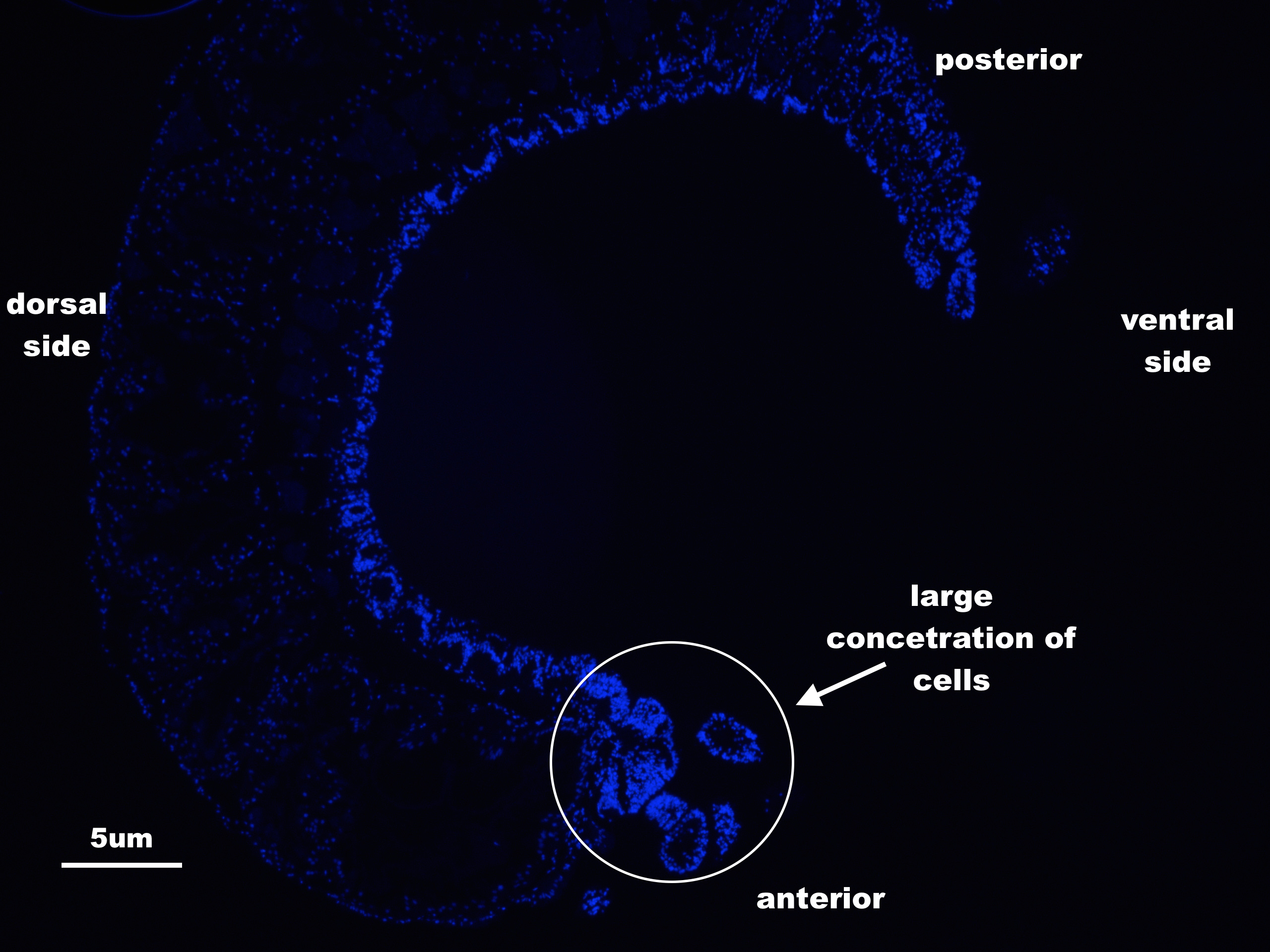
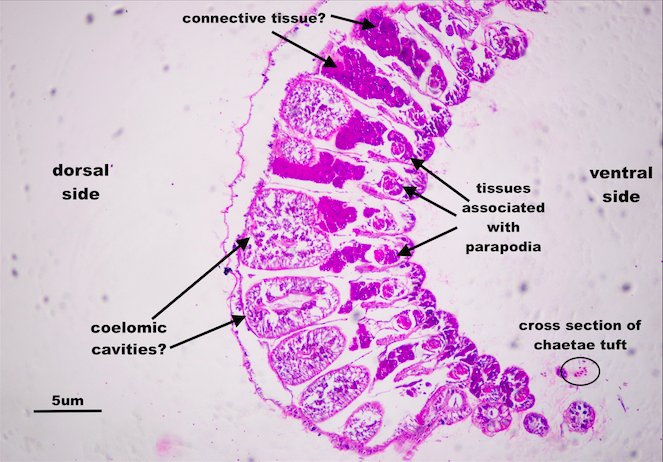
By far the most striking result was the complete absence of any discernible gonads or gametes within the stolons. Not the H&E stained nor the DAPI stained slides revealed any reproductive products (Figure 1). There are two possible explanations for this: 1) that the stolons had not matured enough to start producing gametes, or 2) M. pachycera is included in the group of Autolytinae species that exhibits a gamete ‘production line’. While the first explanation may be credible for some species, the complete lack of gonads in the M. pachycera stolon suggests that this is unlikely. The gamete production line, on the other hand, seems the most plausible explanation.
|
|
Figure 1 Sagittal section of a Myrianida pachycera stolon as seen under a flourescence/brightfield microscope, illuminating cell nuclei blue (top) and tissues and cells pink (bottom). No gonads or gametes were detected. The large concentration of cells at the anterior of the stolon (top) is most likely due to cells associated with ganglion, as stolons tend to have highly developed sensory organs.
|
In Autolytine species such as Autolytus edwarsi, Autolytus prolifera and Autolytus brachycephalus, the female stock has been found to initiate oogenesis inside her own body then, at a particular point in time, transfer the partially developed oocytes to her attached stolon where oogenesis is completed (Siedges, 1979; Franke, 1999). Likewise, male gametes can be produced in Autolytine male stock and transferred to attached stolons (Franke, 1999). Assuming that this gamete production line strategy is exhibited in M. pacycera, it is likely that the examined stolons were still quite young, their gametes yet to be gifted to them.
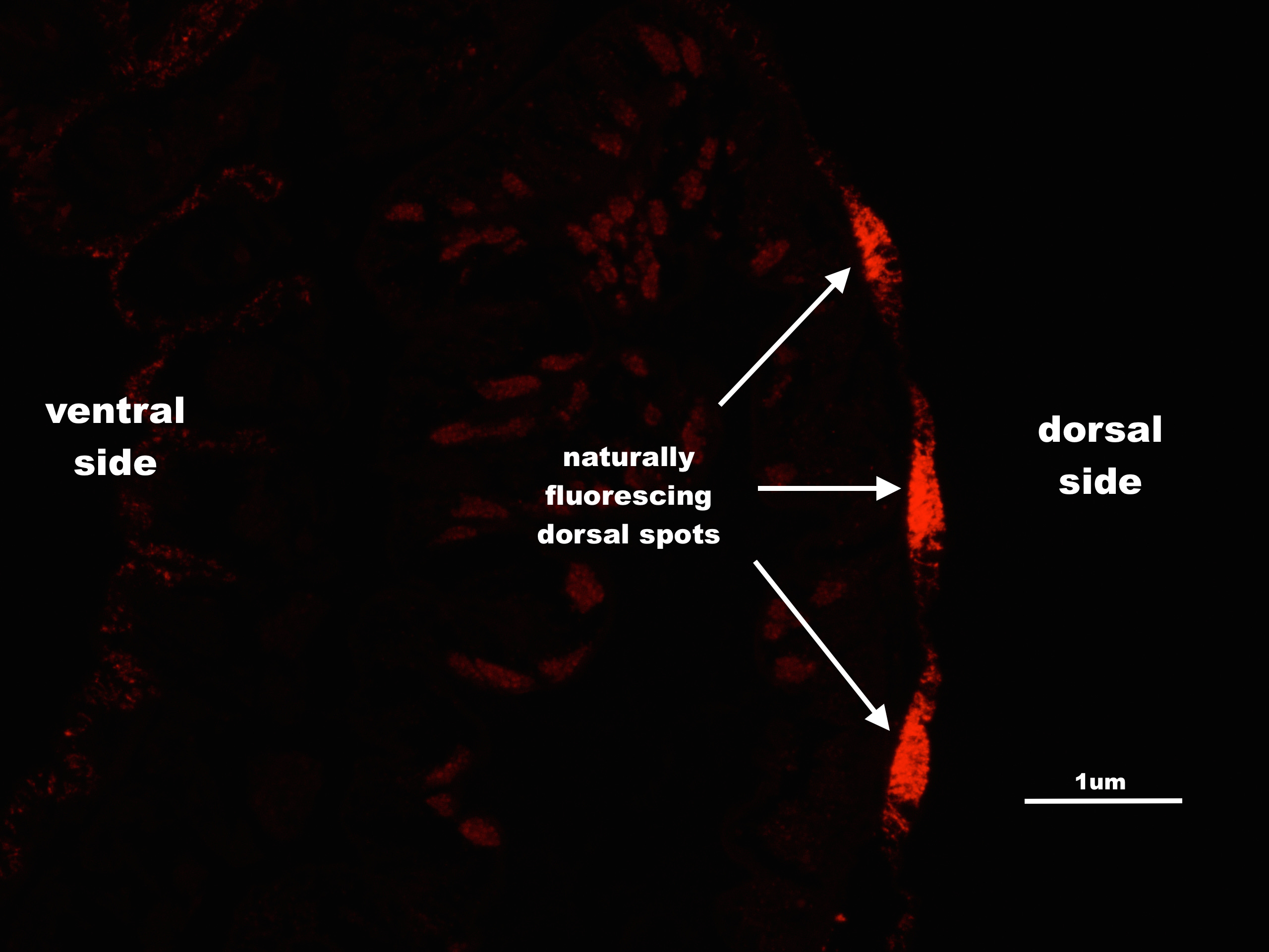
Another interesting finding was the natural fluorescence of the specimen’s dorsal blue spots (Figure 2). There is currently no literature regarding the function, if any, of these spots. But one can’t help but wonder if they and their UV revealed florescence may be implicated in mate recognition. This question was beyond the scope of this study however, but could be considered for future study.
|
|
Figure 2 A stolon of Myrianida pachycera under a fluorescence microscope. Natural fluorescence is illuminated in red.
|
|